













 It’s Only Common Sense: How AI Consumes the Electronics Supply Chain
 Dan’s Biz Bookshelf: ‘Building a Story Brand 2.0’





Phone calls and text messages reach you wherever you are because your phone has a unique identifying number that sets you apart from everybody else on the network. Researchers at the Georgia Institute of Technology are using a similar principle to track cells being sorted on microfluidic chips.
The technique uses a simple circuit pattern with just three electrodes to assign a unique seven-bit digital identification number to each cell passing through the channels on the microfluidic chip. The new technique also captures information about the sizes of the cells, and how fast they are moving. That identification and information could allow automated counting and analysis of the cells being sorted.
The research, reported in the journal Lab on a Chip, could provide the electronic intelligence that might one day allow inexpensive labs on a chip to conduct sophisticated medical testing outside the confines of hospitals and clinics. The technology can track cells with better than 90 percent accuracy in a four-channel chip.
"We are digitizing information about the sorting done on a microfluidic chip," explained Fatih Sarioglu, an assistant professor in Georgia Tech's School of Electrical and Computer Engineering. "By combining microfluidics, electronics and telecommunications principles, we believe this will help address a significant challenge on the output side of lab-on-a-chip technology."
Microfluidic chips use the unique biophysical or biochemical properties of cells and viruses to separate them. For instance, antigens can be used to select bacteria or cancer cells and route them into separate channels. But to obtain information about the results of the sorting, those cells must now be counted using optical methods.
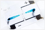
The new technique, dubbed microfluidic CODES, adds a grid of micron-scale electrical circuitry beneath the microfluidic chip. Current flowing through the circuitry creates an electrical field in the microfluidic channels above the grid. When a cell passes through one of the microfluidic channels, it creates an impedance change in the circuitry that signals the cell's passage and provides information about the cell's location, size and the speed at which it is moving through the channel.
This impedance change has been used for many years to detect the presence of cells in a fluid, and is the basis for the Coulter Counter which allowed blood counts to be done quickly and reliably. But the microfluidic CODES technique goes beyond counting.
The positive and negative charges from the intermingled electrical circuits create a unique identifying digital signal as each cell passes by, and that sequence of ones and zeroes is attached to information about the impedance change. The unique identifying signals from multiple cells can be separated and read by a computer, allowing scientists to track not only the properties of the cells, but also how many cells have passed through each channel.
"By judiciously aligning the grid pattern, we can generate the codes at the locations we choose when the cells pass by," Sarioglu explained. "By measuring the current conduction in the whole system, we can identify when a cell passes by each location."
Page 1 of 2