


 Dan’s Biz Bookshelf: ‘Abundance: How We Build a Better Future’
 Trouble in Your Tank: In Complex Systems, Design Rules Aren’t Optional
 It’s Only Common Sense: The Phone Is Still Your Competitive Advantage


New 'Lab-On-A-Chip' Can Test Thousands of Stem Cells Simultaneously
April 10, 2019 | University of ChicagoEstimated reading time: 3 minutes
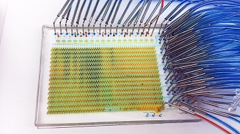
Researchers with the Institute for Molecular Engineering at the University of Chicago have developed a new “lab-on-a-chip” that can examine thousands of individual live cells over a weeklong period, performing experiments that would take more than one million steps in a laboratory.
The credit-card-sized, microfluidic device not only saves time and money, but also offers a new glimpse into how single stem cells react to different molecules and environments.
When researchers examined neural stem cells on the device and analyzed the data, they found several new rules that determine the timing and signaling sequences needed to cause the cells to differentiate or renew themselves. The finding could have implications in understanding brain development or in treating patients with immunotherapy.
“We wanted to develop a microfluidic device that could sort, image and culture single cells in an automated, high-throughput way,” said Assoc. Prof. Savas Tay, lead author of the research, published April 3 in the journal Science Advances. “We achieved that, and now we have an understanding of how stem cells make decisions. That’s pretty exciting.”
Developing a New Way to Study Cells
Cells within our body are constantly responding to different signals and changes in the environment. In stem cells, for example, signals received at different points in time determine how the cell chooses what kind of cell it will develop into. One signal might cause a stem cell to differentiate into another cell, while another signal might cause it to maintain its form.
Researchers currently have no way of studying these signal molecules on individual cells inside the body. Such analysis can be done in a lab with expensive, time-consuming experiments, but they ultimately cannot test all possible outcomes.
Microfluidic devices, which have tiny chambers, tunnels and valves, have offered researchers a faster, automated process for studying these reactions in cells. But these devices have offered a limited number of chambers—meaning researchers could only test a certain amount of conditions with each cell—and could not keep the cells alive long enough to study them over a long period of time.
Finding a Way to Keep Finicky Cells Alive
Tay and his collaborators set out to change that. They designed a microfluidic device that has 1,500 automated chambers—much higher than similar devices, which have less than 100. The device can also conduct several tasks—like cell stimulation, culturing, imaging and sorting—that were previously relegated to separate devices. It can culture cells in different modes—meaning it can examine different types of cells at the same time.
Finally, the device also can keep cells alive for much longer, thanks to a new technique of diffusing media into a cell culture. Normally, to keep cells alive, researchers must change the media they are kept in every few hours. This change shocks the cells, and after several shocks, the cells can die. The researchers’ new technique diffuses the media into the cell chamber, a gentler process that does not shock the cells.
In the first experiment with the device, the researchers studied how different signaling molecules affected the outcome of mouse neural stem cells. Such experiments create millions of data points, so Tay collaborated with Andrey Rzhetsky, UChicago professor in medicine and genetics, to conduct machine-learning analyses on the large dataset.
They found that certain combinations of signals synergize and cause the cells to differentiate, while other molecules shut down those processes. The timing of these signals is also crucial. If a molecule is delivered at the right time, the researchers found, it can change the course of stem cells, from differentiation to self-renewal.
“There are certain orders of signals that are highly optimal, and the exact timing of signals matters,” Tay said. “There hasn’t been a way to dynamically monitor these cells before, so finding and understanding these principles is exciting.”
Next, the researchers hope to use the device to study organoids, tissue cultures derived from stem cells that organize themselves like tiny organs.
Ultimately, a device like this could be used in fields like immunotherapy, where a patient’s own immune system is stimulated to help fight disease. A patient’s stem cells could be removed, placed into the device and be given the right combination of molecules to develop them into a certain lineage, then be placed back in the body.
“We want to be able to use this device for all kinds of problems in cell biology,” Tay said.
Share on:


Subscribe
Stay ahead of the technologies shaping the future of electronics with our latest newsletter, Advanced Electronics Packaging Digest. Get expert insights on advanced packaging, materials, and system-level innovation, delivered straight to your inbox.
Subscribe now to stay informed, competitive, and connected.
Suggested Items
Gregoire Outters Promoted to President of Teledyne Marine Group
04/06/2026 | TeledyneTeledyne Technologies Incorporated announced the promotion of Gregoire Outters to President of its Teledyne Marine Group.
Teledyne Strengthens Commitment to the Space Sector
04/01/2026 | BUSINESS WIRETeledyne Technologies Incorporated is excited to announce the integration of the company’s extensive portfolio of space-focused technologies and businesses, reinforcing its long-term commitment to the global space sector.
Teledyne to Supply Detectors for Lazuli Space Observatory
03/30/2026 | BUSINESS WIRETeledyne Space Imaging, part of Teledyne Technologies Incorporated has been awarded a contract by Schmidt Sciences to deliver advanced near-infrared (NIR) H4RG-10 flight focal plane arrays (FPAs) and custom electronics for integration into the Integral Field Spectrograph on the groundbreaking Lazuli Space Observatory.
New Superconducting Chip Could Enable Breakthrough Nondestructive Terahertz Imaging
03/23/2026 | University of GlasgowA tiny crystal chip which uses terahertz radiation to see clearly through a wide range of materials could find applications in healthcare, biological research, and security screening.
The Test Connection Adds Creative Electron Prime TruVision™ X-ray and CT System for Deeper Failure Analysis
03/05/2026 | TTCIThe Test Connection Inc. (TTCI), a trusted provider of electronic test and manufacturing solutions for more than 45 years, has added the Prime TruVision™ X-ray and computed tomography (CT) inspection system from Creative Electron to its engineering and analysis services.