


 Trouble in Your Tank: Implementing Direct Metallization in Advanced Substrate Packaging
 It’s Only Common Sense: Storytelling That Sells—Stop Pitching, Start Painting Pictures
 The Right Approach: Get Ready for ISO 9001 Version 6


Novel Quantum Dots Enhance Cell Imaging
October 30, 2018 | University of Illinois at Urbana-ChampaignEstimated reading time: 3 minutes
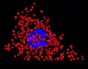
A team of researchers from the University of Illinois at Urbana-Champaign and Mayo Clinic have engineered a new type of molecular probe that can measure and count RNA in cells and tissue without organic dyes. The probe is based on the conventional fluorescence in situ hybridization (FISH) technique, but it relies on compact quantum dots to illuminate molecules and diseased cells rather than fluorescent dyes.
Image Caption: Quantum dots illuminate the locations of individual mRNA as red dots in the cytoplasm of a single HeLa cell. The blue region is the nucleus. This work was a collaborative effort between Illinois Bioengineering and Mayo Clinic researchers.
Over the last 50 years, FISH has evolved into a multi-billion-dollar industry because it effectively images and counts DNA and RNA in single cells. However, FISH has its limitations due to the delicate nature of the dyes. For example, the dyes rapidly deteriorate and are not very good at imaging in three dimensions. In addition, conventional FISH can only read out a couple of RNA or DNA sequences at a time.
"By replacing dyes with quantum dots, there are no stability issues whatsoever and we can count numerous RNAs with higher fidelity than before," said Andrew Smith, an associate professor of Bioengineering and member of the research team. "Moreover, we uncovered a fundamental limit to the size of a molecular label in cells, revealing new design rules for analysis in cells."
In their latest paper, published October 26, 2018, in the online edition of Nature Communications, Smith and his team identified an optimal size for quantum dots in order to effectively work with the FISH protocol. This discovery enabled quantum dot-based FISH to match the labeling accuracies currently obtained with organic dyes.
The team created unique quantum dots that are made of a zinc, selenium, cadmium, and mercury alloy and are coated with polymers. “The core of the dot dictates the wavelength of emission, and the shell dictates how much light will be given off,” said Smith, who is also affiliated with the Micro + Nanotechnology Lab, Carle Illinois College of Medicine, and Department of Materials Science and Engineering at the University of Illinois.
These dots can emit color independent of the size of the particle, which is not the case for conventional quantum dots. The dots are also small enough (7 nanometers) to fit on a probe that can maneuver between proteins and DNA in a cell, making them more comparable in size to the dyes used in conventional FISH probes.
In experiments with HeLa cells and prostate cancer cells, the researchers found that dye-based FISH cell counts declined rapidly in minutes. The quantum dot-based FISH method provided long-term luminescence to allow counting of RNA for more than 10 minutes, making it possible to acquire 3D cell imaging.
"This is important because images of cells and tissues are acquired slice-by-slice in sequence, so later slices that are labeled with dyes are depleted before they can be imaged," said Smith.
This research is part of the Mayo Illinois Alliance in which engineers from Illinois work directly with clinicians and biologists from Mayo Clinic to solve outstanding medical challenges. The Mayo Clinic Biomarker Discovery Group is working to develop FISH-based diagnostics for tumor biopsies in order to improve the accuracy of cancer diagnosis, to select personalized treatments, and to improve prognoses. The QD-FISH methodology was developed to meet this need for biomarker panels requiring the analysis of numerous genetic changes in cells in tumors for which only small amounts of specimens may be available when acquired with a fine needle, as is often the case in prostate cancer.
This research was funded by the Mayo-Illinois Alliance, National Institutes of Health, and the National Science Foundation. The full title of the paper is "Enhanced mRNA FISH with Compact Quantum Dots."
In addition to Smith, the following Illinois Bioengineering researchers contributed to the work: Yang Liu, Phuong Le, Sung Jun Lim, Liang Ma, and Suresh Sarkar. Mayo Clinic collaborators include: Dr. Farhad Kosari, Stephen Murphy, John Cheville, and George Vasmatzis.
Share on:


Testimonial
"The I-Connect007 team is outstanding—kind, responsive, and a true marketing partner. Their design team created fresh, eye-catching ads, and their editorial support polished our content to let our brand shine. Thank you all! "
Sweeney Ng - CEE PCBSuggested Items
MEMS & Imaging Sensors Summit to Spotlight Sensing Revolution for Europe’s Leadership
09/11/2025 | SEMIIndustry experts will gather November 19-20 at the SEMI MEMS & Imaging Sensors Summit 2025 to explore the latest breakthroughs in AI-driven MEMS and imaging optimization, AR/VR technologies, and advanced sensor solutions for critical defence applications.
Direct Imaging System Market Size to Hit $4.30B by 2032, Driven by Increasing Demand for High-Precision PCB Manufacturing
09/11/2025 | Globe NewswireAccording to the SNS Insider, “The Direct Imaging System Market size was valued at $2.21 Billion in 2024 and is projected to reach $4.30 Billion by 2032, growing at a CAGR of 8.68% during 2025-2032.”
I-Connect007’s Editor’s Choice: Five Must-Reads for the Week
07/04/2025 | Marcy LaRont, I-Connect007For our industry, we have seen several bullish market announcements over the past few weeks, including one this week by IDC on the massive growth in the global server market. We’re also closely watching global trade and nearshoring. One good example of successful nearshoring is Rehm Thermal Systems, which celebrates its 10th anniversary in Mexico and the official opening of its new building in Guadalajara.
Driving Innovation: Direct Imaging vs. Conventional Exposure
07/01/2025 | Simon Khesin -- Column: Driving InnovationMy first camera used Kodak film. I even experimented with developing photos in the bathroom, though I usually dropped the film off at a Kodak center and received the prints two weeks later, only to discover that some images were out of focus or poorly framed. Today, every smartphone contains a high-quality camera capable of producing stunning images instantly.
United Electronics Corporation Advances Manufacturing Capabilities with Schmoll MDI-ST Imaging Equipment
06/24/2025 | United Electronics CorporationUnited Electronics Corporation has successfully installed the advanced Schmoll MDI-ST (XL) imaging equipment at their advanced printed circuit board facility. This significant technology investment represents a continued commitment to delivering superior products and maintaining their position as an industry leader in precision PCB manufacturing.