


 Dan’s Biz Bookshelf: Four Important Books You Need to Read (Not Just Say You Have)
 The Marketing Minute: Cracking the Code of Technical Marketing


A Fast Cell Sorter Shrinks to Cell Phone Size
September 24, 2015 | Pennsylvania State UniversityEstimated reading time: 3 minutes
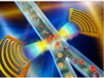
Commercially available cell sorters can rapidly and accurately aid medical diagnosis and biological research, but they are large and expensive, present a biohazard and may damage cells. Now a team of researchers has developed a cell sorter based on acoustic waves that can compete with existing fluorescence-activated cell sorters and is an inexpensive lab on a chip.
"The current benchtop cell sorters are too expensive, too un-safe, and too high-maintenance," said Tony Jun Huang, Penn State professor of engineering science and mechanics. "More importantly, they have very low biocompatibility. The cell-sorting process can reduce cell viability and functions by 30 to 99 percent for many fragile or sensitive cells such as neurons, stem cells, liver cells and sperm cells. We are developing an acoustic cell sorter that has the potential to address all these problems."
Over the past decade, microfluidic cell sorters have emerged as a promising new tool for single cell sequencing, rare cell isolation, and drug screening. However, many of these microfluidic devices operate at only a few hundred cells per second, far too slow to compete with commercial devices that operate on the order of tens of thousands of operations per second. The Penn State system can sort about 3,000 cells per second, with the potential to sort more than 13,000 cells per second.
The researchers achieve the speed by using focused interdigital transducers to create standing surface acoustic waves. When the waves are not focused, the acoustic field spreads out, slowing the sorting process. The narrow field allows the sorting to take place at high speed while gently manipulating individual cells.
"Our high-throughput acoustic cell sorter is expected to maintain cell integrity by preserving not only high viability, but also other cellular features such as gene expression, post translational modification, and cell function," said Huang. "The acoustic power intensity and frequency used in our device are in a similar range as those used in ultrasonic imaging, which has proven to be extremely safe for health monitoring, even during various stages of pregnancy. With the gentle nature of low-power acoustic waves, I believe that our device has the best chance of preserving cell integrity, even for fragile, sensitive cells. Such an ability is important for numerous applications such as animal reproduction, cell immunotherapy, and biological research."
Because the device is built on a lab-on-a chip system, it is both compact and inexpensive -- about the size and cost of a cell phone in its current configuration. With the addition of optics, the device would still be only as large as a book. The researchers fabricated the acoustic cell sorter in Penn State's Nanofabrication Laboratory using standard lithography techniques.
"Just like using a lens to focus light, we design focused interdigital transducers to modify the wave front of acoustic waves and finally confine the waves in a small area, which is comparable with the size of sorting targets," said Liqiang Ren, a graduate student in Huang's group. "The focused acoustic waves have shown better performance in terms of sorting resolution and energy-efficiency than the existing acoustic methods. To the best of our knowledge, our device demonstrates the fastest operation time among all existing acoustic cell sorters."
The researchers, who are from Penn State, Ascent Bio-Nano Technologies and the National Heart, Lung and Blood Institute of the National Institutes of Health, published their work in a recent issue of Lab on a Chip.
"Cell sorting is widely used in many areas of biology to characterize and separate cellular populations of interest," said Philip McCoy, National Heart, Lung, and Blood Institute. "The cytometer size, price, and biohazard concerns remain factors that have prevented this technology from being even more widespread. Microfluidic cell sorting is revolutionary for the fields of cell biology and immunology, as well as other fields in biology, in concomitantly overcoming all of these obstacles. It is quite easy to envision applications for this technology in diverse environments from a family doctor's office to field studies in limnology."
In future work, the researchers plan to integrate their acoustic cell-sorting unit with an optical cell-detecting unit with the goal of increasing throughput to 10,000 events per second.
Additional authors include Yuchao Chen, Peng Li, Zhangming Mao, Po-Hsun Huang, Joseph Rufo and Feng Guo, all of Penn State, Lin Wang of Ascent Bio-Nano Technologies and Stewart J. Levine, National Heart, Lung, and Blood Institute.
The National Institutes of Health, the National Science Foundation, and the Penn State Center for Nanoscale Science supported this work. Portions of the work were performed at the Penn State Nanofabrication Laboratory, a node of the NSF-funded National Nanotechnology Infrastructure Network.
Share on:


Testimonial
"Advertising in PCB007 Magazine has been a great way to showcase our bare board testers to the right audience. The I-Connect007 team makes the process smooth and professional. We’re proud to be featured in such a trusted publication."
Klaus Koziol - atgSuggested Items
MEMS & Imaging Sensors Summit to Spotlight Sensing Revolution for Europe’s Leadership
09/11/2025 | SEMIIndustry experts will gather November 19-20 at the SEMI MEMS & Imaging Sensors Summit 2025 to explore the latest breakthroughs in AI-driven MEMS and imaging optimization, AR/VR technologies, and advanced sensor solutions for critical defence applications.
Direct Imaging System Market Size to Hit $4.30B by 2032, Driven by Increasing Demand for High-Precision PCB Manufacturing
09/11/2025 | Globe NewswireAccording to the SNS Insider, “The Direct Imaging System Market size was valued at $2.21 Billion in 2024 and is projected to reach $4.30 Billion by 2032, growing at a CAGR of 8.68% during 2025-2032.”
I-Connect007’s Editor’s Choice: Five Must-Reads for the Week
07/04/2025 | Marcy LaRont, I-Connect007For our industry, we have seen several bullish market announcements over the past few weeks, including one this week by IDC on the massive growth in the global server market. We’re also closely watching global trade and nearshoring. One good example of successful nearshoring is Rehm Thermal Systems, which celebrates its 10th anniversary in Mexico and the official opening of its new building in Guadalajara.
Driving Innovation: Direct Imaging vs. Conventional Exposure
07/01/2025 | Simon Khesin -- Column: Driving InnovationMy first camera used Kodak film. I even experimented with developing photos in the bathroom, though I usually dropped the film off at a Kodak center and received the prints two weeks later, only to discover that some images were out of focus or poorly framed. Today, every smartphone contains a high-quality camera capable of producing stunning images instantly.
United Electronics Corporation Advances Manufacturing Capabilities with Schmoll MDI-ST Imaging Equipment
06/24/2025 | United Electronics CorporationUnited Electronics Corporation has successfully installed the advanced Schmoll MDI-ST (XL) imaging equipment at their advanced printed circuit board facility. This significant technology investment represents a continued commitment to delivering superior products and maintaining their position as an industry leader in precision PCB manufacturing.