


 Dan’s Biz Bookshelf: Four Important Books You Need to Read (Not Just Say You Have)
 The Marketing Minute: Cracking the Code of Technical Marketing


Microfluidic Devices Gently Rotate Small Organisms and Cells
March 25, 2016 | Pennsylvania State UniversityEstimated reading time: 2 minutes
A method to rotate single particles, cells or organisms using acoustic waves in a microfluidic device will allow researchers to take three dimensional images with only a cell phone.
Acoustic waves can move and position biological specimens along the x, y and z axes, but for the first time researchers at Penn State have used them to gently and safely rotate samples, a crucial capability in single-cell analysis, drug discovery and organism studies.
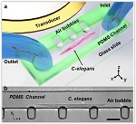
The research, published today in Nature Communications, was led by Tony Jun Huang, professor of engineering science and mechanics and Huck Distinguished Chair in Bioengineering Science. Huang and his group created an acoustofluidic rotational manipulation (ARM) method that traps bubbles in a series of small cavities inside a microfluidic device. Acoustic transducers similar to ultrasound imaging transducers create an acoustic wave in the fluid, making the bubbles vibrate, which creates microvortexes in the flowing liquid that are tunable so the sample rotates in any direction and at any desired speed.
"Currently confocal microscopes are required in many biological, biochemical and biomedical studies, but many labs do not have access to a confocal microscope, which costs more than $200,000," said Huang. "Our ARM method is a very inexpensive platform and it is compatible with all the optical characterization tools. You can literally use a cell phone to do three-dimensional imaging."
To demonstrate the device's capabilities, the researchers rotated C. elegans, a model organism about a millimeter in length frequently used in biological studies. They also acoustically rotated and imaged a HeLa cancer cell.
Existing methods of manipulating small objects depend on the optical, magnetic or electrical properties of the specimen, and/or damage the specimen due to laser heating. The ARM method, on the other hand, uses a gentle acoustic wave generated by a power similar to ultrasound imaging, and at a lower frequency. The device is also compact and simple to use.
"Our method is a valuable platform for imaging and studying the effect of rotation at the single cell level," said co-lead author Adem Ozceki, graduate student in engineering science and mechanics. "More important, with the capacity to rotate large numbers of cells in parallel, researchers will be able to perform high-throughput single-cell studies. "
In addition to its applicability to a large range of biological and physical science investigations, ARM technology shows excellent biocompatibility in a HeLa cell viability test in which 99.2 percent of cells survived manipulation.
Also contributing to "Rotational manipulation of single cells and organisms using acoustic waves" were former group member Daniel Ahmed, Ph.D.; graduate students Nagagireesh Bojanala, Nitesh Nama, Awani Upadhyay, Yuchao Chen; and Wendy Hanna-Rose, associate professor of biochemistry and molecular biology; all from Penn State.
Share on:


Testimonial
"The I-Connect007 team is outstanding—kind, responsive, and a true marketing partner. Their design team created fresh, eye-catching ads, and their editorial support polished our content to let our brand shine. Thank you all! "
Sweeney Ng - CEE PCBSuggested Items
MEMS & Imaging Sensors Summit to Spotlight Sensing Revolution for Europe’s Leadership
09/11/2025 | SEMIIndustry experts will gather November 19-20 at the SEMI MEMS & Imaging Sensors Summit 2025 to explore the latest breakthroughs in AI-driven MEMS and imaging optimization, AR/VR technologies, and advanced sensor solutions for critical defence applications.
Direct Imaging System Market Size to Hit $4.30B by 2032, Driven by Increasing Demand for High-Precision PCB Manufacturing
09/11/2025 | Globe NewswireAccording to the SNS Insider, “The Direct Imaging System Market size was valued at $2.21 Billion in 2024 and is projected to reach $4.30 Billion by 2032, growing at a CAGR of 8.68% during 2025-2032.”
I-Connect007’s Editor’s Choice: Five Must-Reads for the Week
07/04/2025 | Marcy LaRont, I-Connect007For our industry, we have seen several bullish market announcements over the past few weeks, including one this week by IDC on the massive growth in the global server market. We’re also closely watching global trade and nearshoring. One good example of successful nearshoring is Rehm Thermal Systems, which celebrates its 10th anniversary in Mexico and the official opening of its new building in Guadalajara.
Driving Innovation: Direct Imaging vs. Conventional Exposure
07/01/2025 | Simon Khesin -- Column: Driving InnovationMy first camera used Kodak film. I even experimented with developing photos in the bathroom, though I usually dropped the film off at a Kodak center and received the prints two weeks later, only to discover that some images were out of focus or poorly framed. Today, every smartphone contains a high-quality camera capable of producing stunning images instantly.
United Electronics Corporation Advances Manufacturing Capabilities with Schmoll MDI-ST Imaging Equipment
06/24/2025 | United Electronics CorporationUnited Electronics Corporation has successfully installed the advanced Schmoll MDI-ST (XL) imaging equipment at their advanced printed circuit board facility. This significant technology investment represents a continued commitment to delivering superior products and maintaining their position as an industry leader in precision PCB manufacturing.